HEG neurofeedback
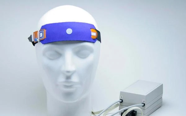
[vc_row][vc_column width=”2/3″][vc_column_text]Two common types of neurofeedback include neurofeedback training through electroencephalographic signals (EEG NFT) and neurofeedback training through hemoencephalographic signals (HEG NFT) or HEG neurofeedback. HEG NFT itself is performed in two ways: NIR-HEG and pIR-HEG
Training to control and manage the physiological functions of the body to achieve the desired state can be achieved through complex means of recording various physiological responses of the body. The term biofeedback describes the process of non-invasive feedback of physiological signals with the aim of establishing control over these signals. Neurofeedback is a subset of biofeedback with the aim of training the brain’s function, either with the aim of improving the symptoms of a specific neurological disorder or with the aim of improving performance using recorded signals from the brain.
NIR-HEG is the first type of HEG introduced by Herschel Thomim (1995). The process involves the use of red light and infrared (infra-red) wavelengths instead of using functional magnetic resonance imaging (fMRI). The second type of PIR-HEG is a system that was later presented by Jeffrey Carman (1997) and the process involves controlling changes in blood flow using infrared light wavelengths through recording changes in the thermal activity of the brain.[/vc_column_text][/vc_column][vc_column width=”1/3″][vc_single_image image=”1545″ img_size=”full” alignment=”center”][/vc_column][/vc_row][vc_row][vc_column][vc_column_text]
Physiological basics:
In both HEG NFT methods, the goal is to quantify brain metabolic activity or the amount of brain metabolism, which is a broad concept in brain imaging science. Metabolism is the burning of glucose in the cell in order to provide the required energy, which is associated with the consumption of oxygen and the production of carbon dioxide, so the amount of metabolism is the amount of energy that is used. So, when the brain is engaged in a mental task, it is expected that it consumes more energy and of course the consumption of oxygen also increases. Although the brain is only 2% of the body weight, it consumes approximately 20% of the available oxygen and 25% of the glucose at rest, and for this purpose, it has a dense network of blood vessels and capillaries that can provide the required oxygen and glucose to the tissue. put a brain We can measure metabolic rate indirectly in different ways, some of these methods are based on a phenomenon called neurovascular coupling. This phenomenon is a mechanism to match the blood flow with the amount of metabolism in the brain. This means that an increase in neural activity in a part of the brain (for example, due to the involvement of the brain in a specific mental task) causes a rapid increase in the speed of blood flow in that particular place, and as a result, the oxygenated blood in this area increases and the oxygen density is high. It is said that this phenomenon is carried out by a type of brain glial cells called astrocytes.
In general, there are two broad categories of brain scanners, one that looks at brain structure and the other that looks at brain activity, which indirectly measures metabolic activity. In particular, the second type of scanners are more commonly used in research to determine which brain area is involved in a specific task, such as PET (positron emission tomography) and SPECT (single photon emission computed tomography), fMRI (functional magnetic resonance imaging) and …
In PET and SPECT methods, glucose consumption is considered as brain fuel. In these methods, glucose marked by radioactive atoms enters the bloodstream and is tracked through the rays emitted from them. In these methods, it is assumed that most of this glucose is sent to a place in the brain where brain activity is higher. In fMRI imaging, blood oxygen is tracked. Increased blood oxygen levels change the magnetic properties of the blood, and this change is detected when the brain is exposed to a strong magnetic field.
In HEG, similar to fMRI, changes in brain activity are detected by detecting changes in blood oxygen density, which can be summarized as follows:
1- A specific mental task activates neurons in some specific areas of the brain.
2- Of course, these neurons consume more energy.
3- This energy demand increases blood flow to the site.
4- Blood oxygen density increases in the place and is tracked by the HEG sensor.
Types of HEG sensors:
As mentioned, there are two types of HEG neurofeedback, each with its own sensors that measure different aspects of the same process, resulting in the same output:
1- NIR-HEG
2- PIR-HEG
 NIR-HEG is historically the first form of HEG and was invented by Dr. Tomim. He presented a method called infrared spectroscopy. Dr. Tomim recognized that the signal he records from this method is continuously affected and its changes can be tracked and therefore useful in the field of biofeedback training.
Comparison of HEG neurofeedback and EEG neurofeedback:
Compared to EEG neurofeedback, the HEG training method has the following advantages:
1- The HEG signal is easier to interpret, the signal only increases or decreases in terms of value, but the EEG signal, in addition to the value, has different frequency bands, each of which can be interpreted.
2- HEG signal is more stable; The EEG signal fluctuates so rapidly that it sometimes appears random.
3- The HEG signal is less exposed to external artifacts.
4- According to Dr. Tomim, from a clinical point of view, through HEG training, we reach treatment goals sooner.
HEG neurofeedback training:
In HEG neurofeedback, one tries to raise the signal. In fact, when the signal increases, the person receives positive feedback and the brain receives reinforcement through the reward system and conditioning takes place. The increase in the HEG signal is caused by the increase in oxygen density in the vessels under the sensor and often results from the increase in the activity of that area. So, for HEG training, the following should be observed (at least for the forehead, which is the most common place for HEG neurofeedback):
1- Loudness and severe mental alertness;
2- motivation and serious intention at the same time to increase and raise the signal;
3- Existence of calmness and positive emotional state (in practice, we must be careful not to create obsession in the person to get the result because researches have proven that the feeling of failure reduces the activity in the frontal cortex).
Application of HEG neurofeedback training:
HEG neurofeedback is a new training method and not many researches have been done on its effectiveness in various disorders, and it may be effective in the treatment of many disorders, and therefore it is an attractive and new area of research in the field of biofeedback.
Based on the researches, HEG has been proven in three areas and has a high effectiveness factor and can be used in treatment:
1- ADD (adult hyperactivity)
2- Depression
3- Migraine
What all three disorders have in common is dysfunction in the prefrontal cortex (PFC). The PFC is a region of the cortex, just behind the frontal bone and above the eyeball, and is known as the executive control center of the brain, and its function plays a central role in goal-directed behavior. PFC plays a significant role in decision-making, planning and execution of programs and maintaining attention to goals in the presence of distracting stimuli. One of the most important functions of the PFC is to coordinate brain resources to execute a goal. Also, the PFC is strongly associated with motivation and excitement. The PFC has the ability to inhibit brain structures involved in emotions. Emotions are related to decision making. The PFC appears to be involved in decision making by visualizing the emotions that may result from each choice. Additionally, this brain region is specifically related to social emotions because the ability to imagine how other people are thinking and feeling is dependent on PFC activity.
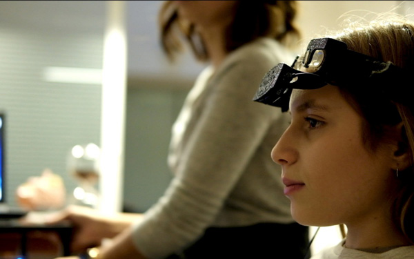
The most common location in HEG neurofeedback training is the PFC region, and most sensors are placed on FP1 and FP2 points according to the 10-20 international system. However, training is possible in all brain locations, and it is possible to examine the effectiveness of HEG in other locations in research based on background and evidence.
Brain mapping studies have shown differential PFC activity in individuals with ADD, often reporting less activity during tasks requiring focused attention. HEG NFT provides guaranteed results for ADD. For this disorder, HEG signal enhancement in the PFC region is often recommended.
Research evidence shows decreased motivation and energy and poor concentration in depressed people. Some depressed people experience emotional monotony and others suffer from intense and painful emotions. All of these can be due to disorganization in the PFC.
Training to increase PFC activity is the best choice for neurofeedback training in depressed people. Firstly, because brain mapping data have reported less activity in this area in depressed people, and secondly, this area plays an important role in other emotional centers of the brain.
Regarding migraine disorder, it can be said that PIR-HEG neurofeedback training has been specifically used for migraine treatment. Dr. Karman was the first to use this technology to treat migraines and treated 100 migraine patients over 4 years and more than 90% of patients who completed at least 6 sessions of PIR-HEG were cured. The neuropathological underpinnings of migraines are not yet definitively understood, but Dr. Carmen’s opinion is that HEG NFTs strengthen the PFC’s inhibitory control over certain parts of the brainstem that appear to trigger migraines.
The effectiveness of HEG in other disorders such as ADHD, anxiety, panic attacks, etc. has also been investigated, but either it was eclectic with other methods, or the number of studies was not enough to be able to express the effectiveness with certainty. did But in general, research has proven that HEG training before training other types of biofeedback can be more effective and shorten the length of training.
References:
Carmen, J.A.(2002). Passive infrared hemoencephalography, 4 years and 100 migraines later: Presented at 2002 society for neuronal regulation conference, Scottsdale, AZ.
Toomim, H., Carmen, J.A.(1999). Hemoencephalography(HEG). Biofeedback, 27(4), 10-14, 27.
Toomim,H.et al.(2004). International increase of cerebral blood oxygenation using hemoencephalography(HEG):An Efficient brain exercise therapy. Neurotherapy, 8(3):5-21; Reprinted in tinius T.(ED.) new developments in blood flow hemoencephalography, Haworth medical press, 2004:5-21.
Sera sala, M.et al.(2012). Evaluating prefrontal activation and its relationship with cognitive and emotional processes by means of hemoencephalography (HEG). Journal of Neurotherapy: Investigations in neuromodulation, neurofeedback and applied neurosciense, 16:3,183-195.
Amen, D.(2003). Healing anxiety and depression
Goldberg, E.(2001). The executive brain: frontal lobes and the civilized mind, oxford university press.putnam.
[/vc_column_text][/vc_column][/vc_row]

